Why Is Medicai the Best for Advanced Visualization Capabilities?
Many radiology teams still wait minutes or hours for large DICOM studies to load because their viewers run locally on individual machines. That delay affects same-day reads, remote consults, and multi-site tumor boards. Medicai places the full viewer in the browser so studies open in seconds from any workstation.
By the end of this article you will know how the zero-footprint viewer, cloud PACS, and AI-supported tools work together, where the platform fits in current workflows, and which care settings benefit most.
What Is Medicai?
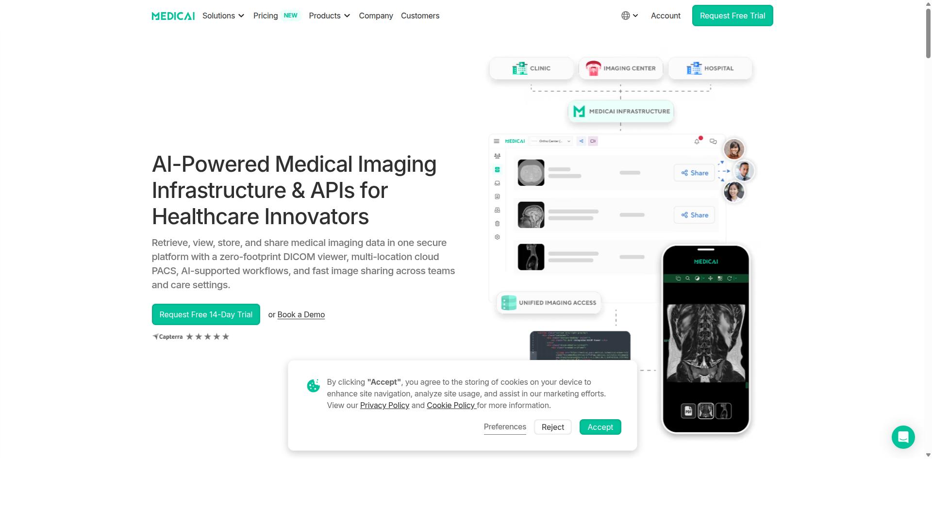
Medicai is a cloud-based medical imaging platform built for healthcare providers that need secure, unified access to imaging studies.
The platform serves as a complete solution for retrieving, viewing, storing, and sharing medical imaging data. A zero-footprint DICOM viewer enables clinicians to access studies through any web browser without installing software.
HIPAA and GDPR compliance protect patient information across every transaction. FDA and CEE clearance confirm the platform meets regulatory standards for medical device software in the regions it serves.
Medicai processes more than 50 million API transactions each year. This high volume demonstrates strong interoperability across different healthcare systems and institutions.
The platform offers Imaging Infrastructure as a Service that modernizes traditional imaging workflows. Healthcare teams gain unified access to imaging studies regardless of their physical location.
Why Medicai Delivers the Best Advanced Visualization Capabilities
This section demonstrates how Medicai's advanced visualization stack improves diagnostic accuracy across CT, MRI, PET, and ultrasound studies.
Advanced visualization transforms raw imaging data into actionable clinical insights. Medicai integrates multiple reconstruction and analysis tools that work together within a unified platform.
Each capability addresses specific diagnostic challenges that radiologists face daily. The system processes studies from multiple modalities without requiring separate software applications.
Medical imaging professionals need tools that deliver both precision and efficiency. Medicai combines traditional visualization methods with quantitative analysis features to support comprehensive patient assessment.
3D Reconstruction
Three-dimensional reconstruction converts cross-sectional images into spatial models that reveal anatomical relationships. Clinicians can rotate, zoom, and section these models to examine structures from optimal angles.
Cardiac surgeons use 3D reconstruction to plan valve replacement procedures. This approach allows precise measurement of annular dimensions before entering the operating room.
Preoperative planning with 3D models reduces procedure time and improves placement accuracy. Surgeons report increased confidence when reviewing spatial relationships that remain hidden in standard planar views.
Multiplanar Reconstruction
Multiplanar reconstruction generates images in arbitrary planes from volumetric datasets. This technique enables viewing of oblique structures that standard axial slices cannot adequately display.
Spine surgeons rely on multiplanar views to assess vertebral alignment and nerve root compression. They can create planes parallel to disc spaces for accurate measurement of herniation extent.
Reformatted images improve communication between radiologists and referring physicians. Treatment decisions become more consistent when all parties examine identical anatomical perspectives.
Volume Rendering
Volume rendering displays three-dimensional datasets with tissue-specific opacity and color assignments. This method reveals internal structures while maintaining spatial context throughout the volume.
Interventional radiologists use volume rendering to map vascular anatomy before catheter placement. The technique highlights vessel origins and branching patterns that guide device selection.
Clear visualization of vascular territories reduces contrast volume requirements during procedures. Patients experience shorter fluoroscopy times when anatomy is mapped accurately beforehand.
Lesion Detection
Lesion detection algorithms identify potential abnormalities across large imaging volumes. These tools flag areas that require closer examination by the interpreting physician.
Chest radiologists apply lesion detection when screening high-risk patients for lung nodules. The system highlights candidates that might otherwise be overlooked during rapid case review.
Standardized detection improves consistency across different readers and experience levels. Departments report fewer missed findings when algorithmic assistance supplements human interpretation.
Tumor Volumetry
Tumor volumetry calculates precise three-dimensional measurements of neoplastic lesions. Serial measurements track treatment response more accurately than traditional diameter assessments.
Oncologists monitor chemotherapy effectiveness through volumetric changes in target lesions. Growth rate calculations inform decisions about continuing, modifying, or switching treatment regimens.
Quantitative tracking replaces subjective size estimates with reproducible measurements. Clinical trials benefit from standardized response criteria that reduce inter-observer variability.
Contrast Enhancement
Contrast enhancement tools adjust image display to highlight differences between tissues. Window level adjustments optimize visualization of specific anatomical structures or pathological processes.
Neuroradiologists apply targeted contrast settings when evaluating subtle brain lesions. Optimized windows reveal enhancement patterns that indicate blood-brain barrier disruption.
Appropriate contrast adjustment improves lesion conspicuity without additional radiation exposure. Diagnostic confidence increases when subtle findings become clearly visible through proper display settings.
Slice Thickness Control
Slice thickness control allows reconstruction of images at varying intervals from source data. Thinner slices provide detail while thicker sections improve signal-to-noise ratios for specific applications.
Abdominal radiologists adjust slice thickness when evaluating liver lesions in different phases of contrast enhancement. Optimal thickness balances spatial resolution against image noise for each clinical question.
Flexible reconstruction reduces the need for repeat acquisitions when initial protocols prove inadequate. Workflow efficiency improves when a single study supports multiple clinical scenarios.
DICOM Tag Editing
DICOM tag editing enables correction of metadata errors that affect image display and routing. Accurate tags ensure proper study identification and maintain compatibility with downstream systems.
Quality assurance teams correct patient identification errors before finalizing reports. Proper tag values prevent misfiling and support accurate longitudinal comparison studies.
Corrected metadata improves interoperability between different vendor systems. Studies transfer successfully across institutional boundaries when standard compliance is maintained.
Quantitative Analysis
Quantitative analysis extracts numerical measurements from imaging data to support objective clinical assessment. These tools calculate parameters such as tissue density, blood flow, and metabolic activity.
Cardiac imagers measure ejection fraction and wall motion from cine sequences. Standardized measurements enable consistent evaluation across different acquisition protocols and equipment vendors.
Objective data replaces subjective visual estimates with reproducible numerical values. Treatment decisions gain precision when supported by quantitative rather than qualitative findings.
Image Fusion
Image fusion combines datasets from different modalities to display complementary information in a single view. PET-CT fusion, for example, overlays metabolic activity onto anatomical structures.
Radiation oncologists use fused images to define treatment volumes that incorporate both tumor extent and functional characteristics. Planning margins become more precise when targeting is informed by multiple data sources.
Fused datasets improve target delineation while reducing geographic miss. Treatment outcomes benefit from accurate localization of disease that single-modality imaging might underestimate.
Performance at Scale
Performance metrics validate that 1 M+ studies are processed yearly without latency spikes.
Medicai's cloud architecture supports 300k+ visualizations per year and maintains 1.7M+ studies in storage. This capacity serves 70 clinics and hospitals with 10,000+ active doctors on the platform.
The system handles 50M+ yearly API transactions while maintaining HIPAA and GDPR compliance. Microsoft Azure partnership provides the infrastructure foundation for consistent performance across all user loads.
Scalable architecture eliminates bottlenecks that affect traditional on-premise solutions during peak demand periods. Users experience reliable access regardless of study volume or concurrent user count.
Global deployment supports real-time collaboration across different time zones and institutions. The 2M+ imaging studies uploaded demonstrate platform capacity that grows with institutional needs.
Zero-Footprint DICOM Viewer and Cloud Architecture
Zero-footprint access eliminates local installations and mitigates security risks for hospitals and imaging centers.
The security model relies on encrypted cloud storage and controlled API access. Medical data security stays centralized while users reach studies through any browser.
Browser-based loading times stay short because images stream on demand instead of downloading entire studies first. Radiologists open cases quickly without waiting for large file transfers.
Integration with existing PACS happens through HL7 and FHIR APIs. The Medicai Imaging API supports over 50 million yearly transactions and proves reliable real-time interoperability across hospital systems.
CONNECT and RETRIEVE services include the Medical Imaging Uploader and DICOM Gateway. These tools feed studies into the Cloud PACS without manual file handling.
STORE and MANAGE offerings cover the Vendor Neutral Archive and Backup and Disaster Recovery. Hospitals keep images accessible while maintaining compliance standards.
ACCESS, VISUALIZE, and COLLABORATE features deliver the DICOM Viewer with real-time collaboration tools. Multiple specialists review cases simultaneously from different locations.
The Doctor Imaging Portal and Patient Imaging Portal extend access beyond radiology departments. Referring physicians and patients view studies through secure web interfaces.
This architecture supports advanced visualization across CT scan, MRI, ultrasound, and PET scan modalities. Multiplanar reconstruction and volume rendering work directly in the browser without additional software.
Structured Reporting for Radiology and AI-Powered Diagnostics integrate into the same workflow. Findings and measurements stay linked to DICOM tags for consistent documentation.
AI-Supported Workflows and Structured Reporting
AI modules automate lesion detection and quantitative measurements, reducing manual steps for radiologists. AI segmentation and annotation tools create structured reports that capture findings in consistent formats.
Medicai integrates AI capabilities from Rayscape.ai and MD.ai partnerships. These integrations bring lesion detection, annotation, and measurement functions directly into the platform workflow.
Radiologists open a CT scan in the viewer. The system identifies potential lesions, calculates volumes, and populates report fields with measurements. Structured reports generate automatically from annotated images.
Neuroaxis reduced time spent reviewing non-relevant cases by 90 percent. The clinic also reported 70 percent fewer face-to-face check-ups while maintaining diagnostic precision across 1,300 cases.
YTS-Dental View achieved 90 percent faster access to patient studies across 10 locations. Both organizations demonstrate how AI-supported workflows improve reporting efficiency without sacrificing accuracy.
Multi-Location Cloud PACS and Collaboration Tools
Cloud PACS links multiple sites, allowing simultaneous review and instant sharing of studies.
Medicai offers a cloud-based platform that connects healthcare facilities across different locations. This setup supports real-time collaboration between radiologists and specialists working at separate sites.
The Standard plan supports one connected location while the Starter plan supports zero. Both plans include access to the core modules that handle medical imaging workflows.
Cloud PACS serves as the central storage system for medical images. It organizes studies from various modalities including CT scan, MRI, ultrasound, and PET scan data.
Patient Case Management organizes patient records and imaging studies into structured cases. Specialists can review complete patient histories with associated images in one location.
Vendor Neutral Archive stores medical images in a format independent of specific equipment manufacturers. This approach ensures long-term accessibility regardless of hardware changes.
Medical Image Exchange facilitates secure transfer of studies between healthcare providers. Authorized users can access and share DICOM files through encrypted connections.
These modules work together to support advanced visualization capabilities across multiple locations. Radiologists gain access to consistent tools for reviewing and analyzing medical images regardless of their physical location.
Pricing and Plans
Two transparent subscription tiers match different organizational storage and connectivity needs. Medicai offers clear monthly billing without hidden fees for both plans.
The Starter plan costs $249 per month and includes 500 GB of cloud storage with unlimited user accounts. This tier supports small to medium teams that need core access to advanced visualization tools for CT scans, MRI studies, and ultrasound imaging without location connectivity requirements.
The Standard plan costs $749 per month and provides 2 TB of cloud storage with unlimited users plus one connected location. Healthcare facilities that require PACS integration and DICOM gateway connectivity benefit from this configuration for streamlined radiology workflows.
Yearly billing delivers 15 percent savings on both tiers. The Starter plan reduces to $209 per month while the Standard plan drops to $639 per month when paid annually.
Enterprise customers receive custom pricing that accommodates multiple connected locations and external sites. Per-study pricing remains available as an alternative billing model for organizations with variable imaging volumes.
A free 14-day trial provides full access to Starter plan features without requiring a credit card. The DICOM gateway setup carries a one-time fee of $1,000 per location for facilities that need direct PACS connectivity.
Trust Signals
Independent certifications and usage numbers substantiate Medicai's security posture. Healthcare organizations need reliable platforms that protect patient data while supporting advanced visualization workflows.
HIPAA and GDPR compliance form the foundation of Medicai's data protection approach. These certifications ensure medical imaging systems meet strict regulatory requirements across different regions.
FDA and CEE cleared viewers demonstrate that the platform meets medical device standards. This clearance process validates the safety and effectiveness of the DICOM viewer technology.
The platform processes 1 million studies yearly and maintains 1.7 million studies in storage. These volume statistics reflect consistent reliability and trust from healthcare providers using the system for routine clinical work.
Microsoft Azure and AWS infrastructure provide the technical foundation for secure operations. Both cloud providers maintain enterprise-grade security standards that support medical data handling requirements.
OWASP security guidelines guide ongoing development practices. This approach addresses common vulnerabilities while maintaining the platform's functionality for medical imaging workflows.
Who Should Use Medicai
Hospitals, imaging centers, and specialty practices handling high volumes of cross-sectional imaging benefit most. These organizations require reliable platforms that manage complex studies efficiently while maintaining diagnostic quality across multiple specialties.
Orthopedics teams rely on Medicai for detailed bone and joint evaluations that support precise surgical planning. Neurology departments use the platform for brain and spine imaging where advanced visualization helps identify subtle lesions and structural abnormalities.
Oncology practices benefit from Medicai when tracking tumor progression over time through consistent measurement tools. Radiologists across all fields gain productivity through streamlined workflows that reduce time spent navigating between different systems.
Cardiology specialists access cardiac imaging with tools designed for functional assessment and vessel analysis. Ophthalmology practices utilize the platform for retinal studies while ob-gyn departments handle fetal imaging and reproductive health documentation.
Pulmonology teams evaluate lung conditions through high-resolution CT scans. Dentistry practices review maxillofacial imaging, and gastroenterology departments examine abdominal studies that require detailed organ visualization.
Medicai processed over 300,000 visualizations last year across these medical specialties. This volume demonstrates the platform's capacity to serve diverse healthcare providers while maintaining performance standards for complex imaging needs.
Final Verdict
Medicai combines zero-footprint access, AI tooling, and scalable pricing into one compliant package. This approach delivers advanced visualization capabilities without requiring local installation or heavy hardware investments. Healthcare teams gain immediate access to sophisticated imaging tools while maintaining strict security standards.
Advanced visualization features enable radiologists to work efficiently with CT scan, MRI, ultrasound, and PET scan data. The platform supports 3D reconstruction, multiplanar reconstruction, and volume rendering for detailed anatomical analysis. These capabilities help improve diagnostic accuracy across various medical imaging workflows.
The system integrates PACS integration and supports DICOM viewer functionality for seamless radiology workflow management. Image segmentation, annotation tools, and measurement tools provide quantitative analysis options. Cross-sectional imaging becomes more accessible when teams can share findings through real-time collaboration features.
AI-assisted imaging tools enhance lesion detection and tumor volumetry processes. Image fusion capabilities allow for better comparison across different scan types. The cloud-based platform maintains HIPAA compliance while supporting interoperability standards including HL7 and FHIR protocols.
Medicai USA
7901 4th St N, STE 300
St. Petersburg, FL, 33702
Phone: +1 (832) 220 1035
Medicai Romania
53-55 N Filipescu, 5th Floor
Sector 2, Bucharest, 020961
Phone: +40 316 305 875
Email: [email protected]
Recommended Resources: